| |
This lecture will describe the new method of Coherent X-ray Diffraction for the study of the structures of smallcrystals, which takes advantage of the relatively high coherence of the latest synchrotron X-ray sources. A recentcrystallographic study of a monodispersed Au101RS44 gold nanoparticle-thiol complex revealed a crystallinecore particle coated with a shell of 1nm thickness with enlarged Au-Au spacings and interpenetration of the thiolligand species. Far from having a well-defined boundary between the metal and the organic sides of the interface,this unusual complex was found to contain a mixed compound layer as its lowest energy configuration [1]. Weinvestigated the structure of facetted gold nanocrystals before and after coating with propane thiol, C3H7SH, one ofthe simplest SAM-forming organic molecules. We found our gold nanocrystals to be spherical with {111} facets, asexpected from the theory of Equilibrium Crystal Shapes (ECS). The structure of a single 300nm-diameter particlewas measured using the powerful technique of Coherent X-ray Diffraction (CXD), which is highly sensitive to thepattern of internal strains within the nanocrystal [2]. Our results show that the strain is profoundly modified by thethiol adsorption. We are able to establish the magnitude of this stress difference using finite element calculations andsuggest a model involving preferential reaction on the curved regions of the crystal surface [3].
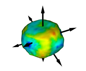 Figure showing the shape of the crystal and the phase found onthe surface of the particle before dosing on a colour scale from -1(blue) to +1 radian (red). The arrows indicate the {111 } directions;the substrate surface normal direction is parallel to the large facetat the top of the figure. The (111) Q-vector used for imaging is onthe right hand side. The phase is the projection of the crystal'sdisplacement field onto this direction [2].
[1] "Structure of a thiol monolayer-protected gold nanoparticle at 1.1 angstrom resolution" P. D. Jadzinsky, G. Calero, C. J. Ackerson, D. A. Bushnell and R. D. Kornberg, Science 318 430-3 (2007)
[2] "Coherent Diffraction Imaging of Strains on the Nanoscale", I. K. Robinson and R. Harder, Nature Materials 8 291-298 (2009)
[3] "Shape-dependent Thiol-induced strains in gold nanocrystals", Moyu Watari, Rachel McKendry, Manuel Vögtli, Gabriel Aeppli, Yeong-Ah Soh, Xiaowen Shi, Gang Xiong, Ross Harder and Ian Robinson in preparation
|